
Peripheral Neuropathy
Reduced sensation may prevent pressure, heat or injury from being noticed. Neuropathy can also change foot mechanics and create areas of excessive pressure.
Specialized diabetic foot care
Comprehensive vascular assessment and individualized treatment planning to control infection, restore blood flow when necessary, support wound healing and preserve functional tissue.
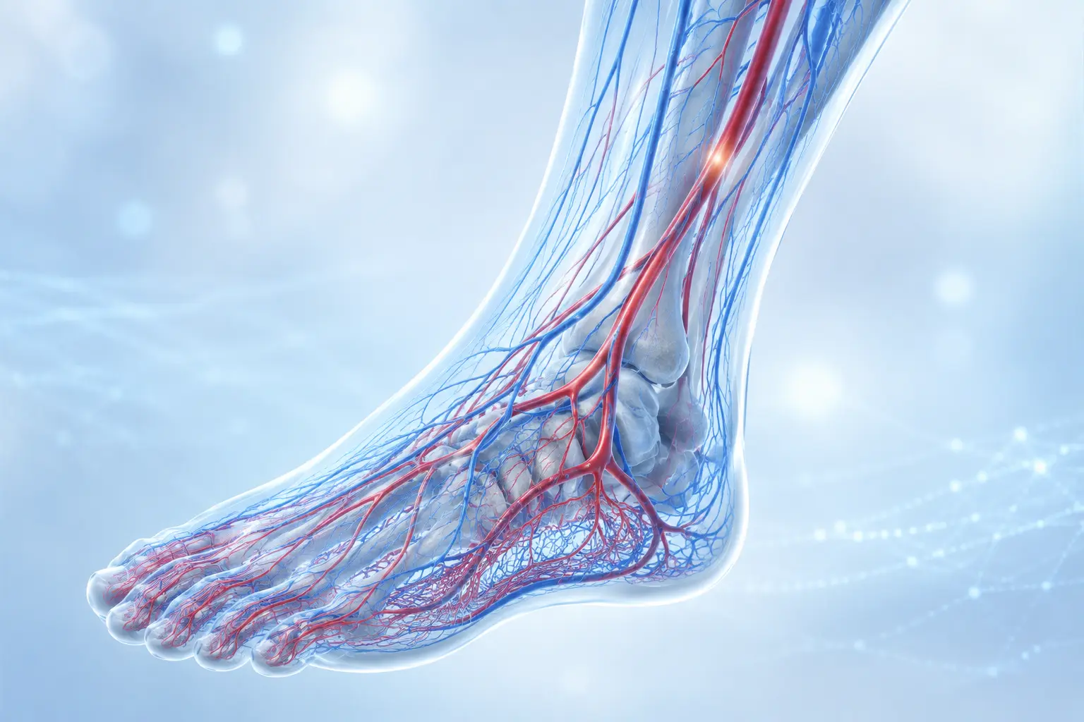
Understanding diabetic foot disease
Diabetes can affect the nerves, blood vessels, skin and natural healing processes of the feet. Even a small blister or pressure point may develop into a serious wound if it is not recognized early. Care is therefore coordinated across vascular surgery, diabetology, wound care, podiatry, infectious diseases, radiology and other disciplines when appropriate.
How problems develop
Reduced sensation may prevent pressure, heat or injury from being noticed. Neuropathy can also change foot mechanics and create areas of excessive pressure.
Narrowed or blocked arteries limit the oxygen and nutrients needed for healing and infection control. Significant arterial disease may be present without obvious symptoms.
An open wound can allow infection to spread into deeper tissues, joints or bone—particularly when circulation is reduced or diabetes is poorly controlled.
Foot deformity, unsuitable footwear and repetitive pressure can cause wounds. Without effective pressure relief, ulcers may fail to heal or return after closure.
Conditions we assess and treat
Ulcers may develop through neuropathy, pressure, poor circulation, injury, infection or a combination of factors. Prompt vascular assessment is important when reduced blood flow may be limiting healing.
Increasing redness, warmth, swelling, discharge, odour or tissue discolouration may indicate infection. Serious infection can be present without significant pain in patients with neuropathy.
A deep or long-standing ulcer may extend into bone. Diagnosis can require clinical assessment, blood tests, X-rays, MRI or a bone sample in selected cases.
Tissue may die because of critically reduced blood flow, severe infection or both. Early assessment helps determine whether circulation can be improved and viable tissue preserved.
A warm, swollen or changing foot shape in a person with neuropathy requires prompt assessment, even when there is little or no pain.
Previous ulceration, amputation, severe neuropathy, foot deformity or peripheral arterial disease increases future risk and requires structured surveillance and preventive care.
Comprehensive assessment
The wound, circulation, sensation, infection risk and foot mechanics are assessed together so that treatment can be prioritized safely.
The history of the wound and relevant health factors are reviewed.
Both feet are checked for wounds, pressure points, callus, infection, deformity, tissue loss, colour and temperature differences.
Sensation and blood supply may be assessed using pulses, Doppler, ankle or toe pressures, transcutaneous oxygen measurements and duplex ultrasound.
Location, size, depth, tissue quality, drainage and signs of infection are documented. Microbiological samples may be taken when clinically useful.
X-rays or MRI can investigate deep infection. Duplex ultrasound, CT or MR angiography and selected catheter angiography can define arterial disease and guide treatment.
The limb-salvage approach
Limb salvage is more than avoiding amputation. A plan may combine several coordinated steps. In advanced situations, major amputation may still be the safest option when infection cannot be controlled, damage is irreversible or repeated treatment would be unsafe.
Control of infection
Restoration of adequate blood flow
Removal of non-viable tissue
Protection and preparation of the wound
Pressure relief
Reconstruction when appropriate
Rehabilitation and preventive follow-up
Treatment journey
The wound, circulation, sensation, infection risk and foot structure are assessed. Urgent problems are identified and prioritized.
Vascular tests, wound investigations and imaging are arranged according to the clinical findings.
The treatment sequence may combine infection control, offloading, wound care, restoration of circulation and surgical care.
Healing is reviewed regularly. After closure, footwear, pressure protection, foot surveillance and risk-factor control remain important.
Prevention
Because neuropathy may mask pain, regular inspection and early assessment of any change remain essential.
Check both feet and between the toes every day
Wear suitable footwear and inspect shoes before use
Avoid walking barefoot
Protect the feet from heat and cold
Keep skin clean and appropriately moisturized
Do not self-treat calluses, corns or ingrown nails
Attend regular diabetic and vascular reviews
Follow individualized footwear and offloading advice
Seek early assessment for blisters, cracks or wounds
Ask a caregiver for help when vision or mobility is limited
Frequently asked questions
The correct treatment depends on circulation, infection, pressure, tissue damage and the patient’s overall health.
No. A wound may be caused by neuropathy, pressure, injury, infection, reduced circulation or several factors together. A complete assessment identifies the main causes.
Diabetic neuropathy can reduce or remove protective sensation. The absence of pain does not mean that a wound is minor or safe to ignore.
No. Vascular treatment is considered when reduced blood supply contributes to poor healing or threatens the limb. Other patients may primarily need offloading, wound care or infection treatment.
In suitable patients, arteries may be treated using balloon angioplasty, sometimes with a stent. Other patients may benefit more from bypass surgery. The decision depends on anatomy and the overall clinical situation.
Antibiotics treat infection but do not correct pressure, dead tissue or poor circulation. Successful treatment may require several coordinated measures.
No treatment can guarantee limb preservation. Early assessment and coordinated care can improve the opportunity for limb salvage, but some severely damaged or infected limbs cannot be safely preserved.
Yes. Recurrence is possible when neuropathy, pressure, deformity or vascular disease remains. Long-term footwear, surveillance and preventive care are essential.
When to seek assessment
The information on this page is intended for general patient education and does not replace an individual medical assessment.